Congenital Myopathies: A Comprehensive Guide on Genetic Muscle Disorders in Children

Overview
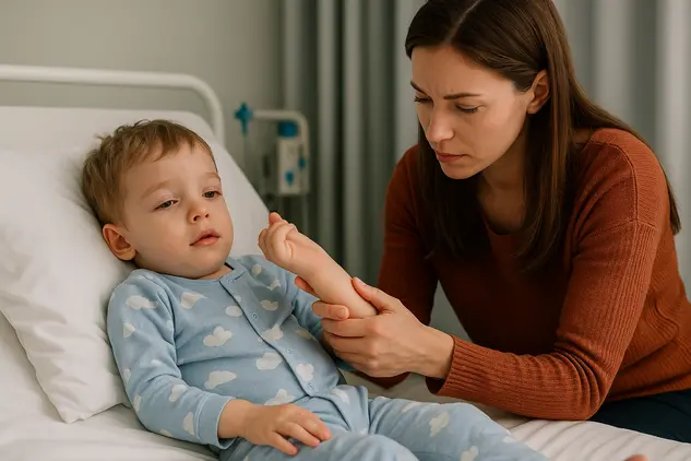
Congenital myopathies are a group of rare, inherited muscle disorders that appear at birth or in early childhood. They affect how muscle fibers are built, structured, or function, leading to low muscle tone (hypotonia), weakness, and delays in motor milestones. While the word congenital suggests “present at birth,” some children present later in infancy or early childhood with milder signs that were not recognized initially.
Unlike muscular dystrophies—which involve progressive muscle fiber breakdown and often rising muscle enzyme levels—many congenital myopathies are characterized by structural abnormalities within the muscle fibers and may have stable or slowly progressive courses. The severity ranges widely. Some infants have significant breathing or feeding challenges early on, while others experience mild motor delays and live active lives with appropriate supports. Early recognition, genetic diagnosis, and tailored care plans can make a substantial difference in health, development, and quality of life.
This guide explains the types, causes, symptoms, diagnosis, treatment options, daily management, and long-term outlook of congenital myopathies. It is designed to help parents, caregivers, educators, and clinicians better understand these conditions and support children with confidence and compassion.
What Is Congenital Myopathy?
Defining congenital myopathy
Congenital myopathy is an umbrella term for genetic muscle conditions characterized by distinctive structural changes visible under the microscope or detected through genetic testing. The hallmark is impaired muscle fiber function due to abnormalities in proteins responsible for contraction, energy handling, or structural stability. Symptoms typically include low muscle tone, muscle weakness, and delayed motor milestones (such as rolling, sitting, or walking).
How congenital myopathy differs from muscular dystrophy
Congenital myopathy:
- Often stable or slowly progressive
- Muscle enzymes (like CK/CPK) are usually normal or only mildly elevated
- Defined by structural abnormalities such as rods, cores, or central nuclei
Muscular dystrophy:
- More commonly progressive muscle degeneration
- Marked elevation in CK/CPK is typical
- Often involves ongoing muscle fiber breakdown and replacement with fat/connective tissue
Who is affected
Congenital myopathies affect boys and girls worldwide and occur across all ethnic backgrounds. Prevalence varies by type and population. Some forms are extremely rare; others, such as RYR1-related myopathies, are among the more frequently diagnosed.
Types of Congenital Myopathy
Nemaline myopathy
- Characterized by rod-like structures (nemaline bodies) in muscle fibers.
- Common genes: NEB (nebulin), ACTA1, TPM2, TPM3.
- Presentation ranges from severe neonatal weakness and respiratory difficulties to milder childhood weakness affecting facial, neck, and limb muscles.
Central core disease (RYR1-related)
- Defined by central “cores” seen on muscle biopsy, indicating areas of reduced oxidative activity.
- Most often caused by mutations in the RYR1 gene, which also confers risk of malignant hyperthermia with certain anesthetics.
- Symptoms vary from mild motor delays to significant weakness, with hip girdle involvement and predisposition to orthopedic issues such as hip dislocation.
Multiminicore disease (SEPN1/SELENON and others)
- Multiple small cores in muscle fibers.
- Often due to SELENON (formerly SEPN1) variants.
- Tends to feature axial muscle weakness (neck and trunk), early scoliosis, and respiratory insufficiency that may be more pronounced than limb weakness.
Centronuclear/myotubular myopathies
- Noted for centrally located nuclei in muscle fibers.
- Genes include MTM1 (X-linked; typically affects boys), DNM2, BIN1, RYR1, and others.
- The X-linked form (MTM1) may cause severe neonatal weakness and breathing challenges; dominant forms (e.g., DNM2) can be milder and present later.
Congenital fiber-type disproportion (CFTD)
- Predominance of smaller type 1 fibers relative to type 2 fibers.
- Can be associated with ACTA1, TPM2/3, RYR1, and other genes.
- Presents with generalized weakness and hypotonia; facial and bulbar involvement vary.
Other and overlapping myopathies
- MYH7-related myopathy
- KLHL40/KLHL41-related severe neonatal myopathy
- TTN-related congenital myopathy
- Overlaps with congenital myasthenic syndromes and metabolic myopathies can occur, highlighting the need for careful diagnosis.
Causes and Genetics
The role of muscle proteins
Muscle fibers rely on a complex network of proteins to contract and maintain structural integrity. Genes like RYR1, NEB, ACTA1, and MTM1 encode proteins involved in calcium release, thin/thick filament assembly, and membrane trafficking. Changes in these genes can disrupt muscle function, leading to low tone and weakness.
Inheritance patterns
Autosomal recessive: Two nonworking copies (one from each parent) are required. Parents are typically carriers without symptoms. Each child has a 25% chance of being affected.
Autosomal dominant: One altered copy is enough to cause disease. Can be inherited from an affected parent or occur de novo (new in the child).
X-linked: The gene is on the X chromosome (e.g., MTM1). Typically, boys are affected; mothers may be carriers with minimal or no symptoms.
Genetic variability and expressivity
Even with the same gene change, severity can differ among family members. Modifying genes, environment, and chance all influence how the condition appears and progresses. This variability underscores the importance of individualized care.
De novo mutations
Some children develop congenital myopathy due to a new genetic change not present in either parent. In such cases, the recurrence risk in future pregnancies may be lower but is not zero because of rare germline mosaicism. Genetic counseling can address specific risks.
Signs and Symptoms
Early signs in newborns and infants
- Hypotonia (floppy baby)
- Weak cry and limited spontaneous movement
- Feeding difficulties (poor suck/swallow), risk of aspiration
- Breathing difficulties, especially during sleep; recurrent respiratory infections
- Delayed motor milestones (head control, rolling, sitting)
Symptoms in toddlers and older children
- Generalized or proximal muscle weakness (hips, shoulders)
- Gait differences (waddling, toe walking), frequent falls
- Difficulty running, jumping, or climbing
- Facial weakness (open mouth posture), drooling, speech clarity challenges
- Joint contractures or hypermobility, depending on type
- Scoliosis due to trunk weakness
Respiratory and sleep-related issues
- Shallow breathing, weak cough, and difficulty clearing secretions
- Sleep-disordered breathing (hypoventilation), morning headaches, daytime sleepiness
- Vulnerability to respiratory infections and complications
Feeding, swallowing, and nutrition
- Poor weight gain, fatigue during feeding, prolonged meals
- Gastroesophageal reflux, constipation
- Risk of aspiration leading to pneumonia if swallowing is unsafe
Less common or condition-specific features
- Eye movement limitations (ophthalmoplegia) in centronuclear myopathies
- Hip dislocation in central core disease
- Cardiac involvement is uncommon overall but can occur in specific subtypes (e.g., MYH7-related)
How Congenital Myopathy Is Diagnosed
Medical history and examination
A pediatric neuromuscular specialist will review prenatal, birth, and developmental histories, family background, and current symptoms. The exam assesses tone, strength, reflexes, cranial nerve function (including facial and bulbar muscles), posture, joint range of motion, and respiratory status.
Laboratory testing
- Creatine kinase (CK/CPK): Often normal or mildly elevated in congenital myopathies.
- Basic labs to evaluate nutrition and rule out metabolic conditions.
- Genetic panels or exome/genome sequencing to identify causative variants.
Electrophysiology and imaging
- Electromyography (EMG) and nerve conduction studies can show myopathic patterns but may be normal; interpretation in children requires expertise.
- Muscle MRI can reveal characteristic patterns of muscle involvement that guide genetic testing and minimize need for biopsy.
Muscle biopsy
A small sample under local or general anesthesia may be examined for rods, cores, central nuclei, or fiber-type disproportion. With advances in genetic testing, biopsies are now less frequently required but remain valuable in select situations.
Differential diagnosis
- Spinal muscular atrophy (SMA): Lower motor neuron disease with denervation; genetic testing distinguishes SMA.
- Congenital myasthenic syndromes: Impaired neuromuscular transmission causing fatigable weakness; respond to specific drugs.
- Muscular dystrophies: Progressive weakness with higher CK.
- Metabolic myopathies, central hypotonia due to brain disorders, and connective tissue disorders.
Newborn screening and carrier testing
Routine newborn screening typically does not include congenital myopathies. If a family mutation is known, prenatal testing or preimplantation genetic testing may be options. Carrier testing can help family planning.
Treatment and Management
No single cure, but many effective supports
There is no universal cure for congenital myopathies yet. However, targeted therapies, supportive care, and proactive monitoring can significantly improve mobility, growth, breathing, comfort, and participation in daily life.
Multidisciplinary care team
- Pediatric neuromuscular specialist (neurologist or physiatrist)
- Pulmonologist/sleep medicine specialist
- Physical therapist, occupational therapist, speech-language pathologist
- Dietitian, gastroenterologist
- Orthopedist and spine specialist
- Genetic counselor, social